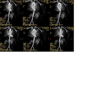
Although no standardized techniques exist for helical CT evaluation of the thoracic aorta, and practices vary from institution to institution, certain general principles may be applied to optimize techniques. Variables that must be considered include collimation, pitch, field-of-view, reconstruction increment, amount, rate and timing of contrast administration, distance to be scanned, kVp, mA, and tube rotation time. These variables must be optimized to provide the highest possible scan quality. Occasionally, a compromise among these parameters is required to achieve this goal.
Collimation and Pitch
When a scan protocol is constructed, one must generally strike a compromise between the collimation and the pitch. Recall that pitch reflects the ratio of table speed to slice thickness (collimation) per tube rotation, or:
Pitch = | Table transport distance Collimation |
A useful general rule of thumb is that effective slice thickness increases by 30% when the pitch is doubled. Thus, a 3-mm scan with a pitch of 2 has an effective slice thickness of 3.9 mm. Apply this concept to the following situation:
What technical parameters provide the best scan quality if the distance to be scanned is 10 cm? If one uses 5-mm collimation with a pitch of 1, the scan will take about 20 seconds (depending on tube rotation time) with an effective slice thickness of about 5 mm. If 3-mm collimation with a pitch of 2 is employed, the scan will require just under 17 seconds with a 3.9 mm effective slice thickness. Thus, whenever possible, it is preferable to maximize spatial resolution using thinner collimation while increasing the pitch to cover the entire scan distance more expediently. a faster scan will increase scanner throughput and minimize respiratory motion degradation.
Field-of-View
The field-of-view (FOV) deserves special consideration for two reasons. First, because the conditions that prompt helical CT examination of the aorta are often nonspecific, evaluation of the entire thorax is important. the diagnosis may be found in the pleural space or peripheral lung parenchyma, not the aorta. Additionally, using small FOVs will decrease photon flux in the imaging volume, contributing increased image noise. This may be partially compensated for by increasing the mA, but the scanner may not permit a sufficient increase in ma if the length of the scan is long. for these reasons, it is advisable to measure the FOV from outer rib to outer rib at the widest portion of the thorax. This FOV creates a good compromise between a large enough FOV to visualize the entire thorax yet provides a larger image with improved resolution that is easier to read.
Reconstruction Increment
A reconstruction increment that provides nearly a 50% overlap in slice thickness is generally sufficient to generate excellent quality images. Recent work suggests that there is limit at which overlapping the reconstructions no longer provide improved longitudinal resolution.[1] Beyond this point, further overlap only generates more images and thus only increases scan reconstruction time and storage requirements. the optimal overlap depends on the pitch employed. As a compromise, a 50% overlap generally provides optimal resolution and enhances the quality of postprocessing techniques. One should avoid using reconstruction increments that are exact harmonics of the collimation employed (i.e., a 2.5-mm reconstruction increment with a 5-mm collimation scan). in such circumstances, artifacts may be introduced if post-processing techniques are used.
Iodinated Contrast
Amount and Concentration: There is no consensus regarding what iodine concentration should be routinely employed for helical CTa of the aorta. the common practice of using undiluted (300 mg I/mL or 360 mg I/mL) is usually sufficient. in one study, an iodine concentration of 125 mg I/mL was found to be unacceptable at any flow rate.[2] in this same study, an iodine concentration of 150 mg I/mL provided satisfactory images, when the flow rate was adjusted. This iodine concentration had the added benefit of decreasing the amount of streak artifact emanating from thoracic venous structures. However, questions regarding the safety of diluted contrast preparations remain because such dilute mixtures are not generally commercially available. Obviously, there are issues of practicality as well. Generally, we employ undiluted contrast (300 mg I /mL) for all thoracic helical CTa applications.
Rate: There is considerable variation among investigators regarding the rate at which contrast is delivered for helical CTa of the thorax. Generally speaking, a rate of 3 cc/sec is sufficient, although some investigators use rates as high as 5 cc/sec. Some consider higher rates as less practical because they may require larger bore catheters; however, rates of 4 to 5 cc/sec may be achieved using 20-gauge catheters. Generally, an injection rate of 3.5 cc/sec is satisfactory for helical CTa applications in the chest. for such injection rates, 22-gauge catheters are adequate. Indeed, flow rates of 4 to 4.5 cc/sec may be used through such catheters when required. It should be noted that when higher injection rates are used, a larger volume of contrast must be delivered to maintain the injection throughout the scan acquisition.
Injection Timing: An appropriate scan delay is critical for excellent vascular imaging. As a general rule of thumb, a standard delay of 25 seconds for helical CTa of the thoracic aorta will usually suffice. Patients with cardiomyopathy or an unsuspected stenosis of the injected vein may require longer delays to achieve optimal aortic opacification. Patients with hyperdynamic cardiac function may require a shorter delay. the time to peak aortic opacification from patient to patient varies considerably. Van Hoe et al[3] found that this delay varied from 11 to 30 seconds, with an average of 20 seconds. One may perform a test injection to determine the time to optimal aortic opacification in a given patient. With this method, a 20-cc bolus of contrast is administered, followed by a 10 second delay. After the 10 second delay, one image every 2 seconds is acquired at the same level (often the cranial aspect of the descending thoracic aorta) for a total of 30 seconds. the image with the greatest contrast density is used to select the proper scan delay. This method is somewhat cumbersome to perform routinely, and has the added detraction of an increased radiation dose as well as a slight increase in visceral background attenuation due to the injected contrast.
As an alternative, bolus timing software packages may be purchased for most modern CT scanners. Such programs allow the time to peak enhancement to be monitored without a separate test injection. These programs monitor the attenuation of the vessel in question during the contrast injection, and display the attenuation graphically in real-time. Once the graph demonstrates a sharp rise in attenuation, the scan sequence is triggered manually. When possible, we prefer to use bolus timing software to ensure proper arterial opacification. With even minimal experience, the CT technologist can learn to achieve excellent arterial opacification without the direct super-vision of the radiologist.
Distance to Be Scanned
The entire distance to be scanned is important for several reasons. Perhaps, most importantly, the contrast injection must be maintained for all but the last few seconds of the scan acquisition. Failure to do so may allow unopacified blood to enter the imaging volume, thus creating artifacts and diminishing the quality of the study. Second, the longer the distance to be scanned, the greater the heat loading on the tube, necessitating a lower ma be used. This is particularly problematic in larger patients, who may require all the photons the tube can muster. Third, imaging beyond the desired structures will provide additional information, but at the expense of more tube exposures. As the x-ray tube has a finite life span, extra tube exposures may come at significant cost. Finally, imaging beyond the desired volume also contributes to increased patient radiation dose.
kVp and mA
There is usually no reason to adjust the kVp from the standard values of 120 to 140 kVp. However, the ma used is critically important for achieving excellent quality studies. While it is preferable to keep ma as low as possible to reduce patient dose, little is accomplished if the patient receives a somewhat lower radiation dose at the expense of a poor quality study. the contrast resolution on CT scanners is heavily dependent on mA. Noisy images resulting from increased quantum mottle often result from using ma values that are too low for a particular patient. Although for the average patient, ma is often not a major consideration, for larger patients, an upward adjustment in the ma is critical to achieve a high quality study. Often this adjustment must be performed manually because modern CT scanners are often programmed to minimize the ma to reduce tube heat loading and patient dose. When larger patients are being scanned, the ma should be adjusted manually to near maximum to diminish image noise and provide the highest quality imaging. Careful attention to FOV and scanning distance in this circumstance is also required.
Tube Rotation Time
Modern scan-ners now allow tube rotation times as low as 0.5 seconds. GE scanners (GE Medical Systems, Milwaukee , WI


