Protocol:
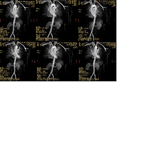
H/O Psot oprative left Common iliac and abdomenal aorta 1.5Tesla.
Contrast Enhanced Angio performed with 3D TOF (Time of flight) sequence. The study viewed in row as well as 3D reconstructed images. The procedure performed in single session with I.V. 25cc Gadolinium (Multihance). MIP images of each side were produced separately to prevent superimposition on the lateral projections. Mask images obtained followed bolus chasing, the rapid moving-table technique used.
1st slab covering thoraco abdominal aorta at the level of diaphragm to femorals up to the level of inguinal ligament , 2nd slab covering lower limb major arteries femoral at the level of inguinal ligament to level of knee joint and 3rd slab covering free knee downward
Contrast-enhanced MR angiography of abdominal aorta and lower extremityH/O Psot oprative left Common iliac and abdomenal aorta 1.5Tesla.
Contrast Enhanced Angio performed with 3D TOF (Time of flight) sequence. The study viewed in row as well as 3D reconstructed images. The procedure performed in single session with I.V. 25cc Gadolinium (Multihance). MIP images of each side were produced separately to prevent superimposition on the lateral projections. Mask images obtained followed bolus chasing, the rapid moving-table technique used.
1st slab covering thoraco abdominal aorta at the level of diaphragm to femorals up to the level of inguinal ligament , 2nd slab covering lower limb major arteries femoral at the level of inguinal ligament to level of knee joint and 3rd slab covering free knee downward